Flash Photography
Los Alamos scientists pioneered techniques to photograph extremely fast phenomena such as implosions.
- Rebecca McDonald, Science Writer
During the Manhattan Project, Los Alamos scientists needed a diagnostic tool. Like an x-ray confirming a broken leg bone, the researchers wanted to be able to see into the smoke cloud of detonation products. They needed to verify that the implosion remained symmetric to ensure an efficient detonation. Although it was a time when scientific breakthroughs came quickly, this challenge stumped them, and their initial efforts to use x-rays to visualize the event were limited. According to Manhattan Project historian David Hawkins, “These objectives were pursued relentlessly, but because of the great technical difficulties involved, with little real success. The program was dropped in March 1945.”
“I find this quote very interesting because it shows just how difficult it is to create these dynamic images,” says Scott Watson, Laboratory Fellow, who has, over his 38-year career, taken hundreds of images of explosions. Despite the setback in 1945, Los Alamos scientists went on to pioneer several capabilities to visualize and evaluate dynamic events, leading to key elements of today’s national security mission.
Can you see it?
Scientists during the Manhattan Project designed the first atomic device as a sphere of high explosives with a metal core. When the high explosives were detonated, they would implode and compress the metal core symmetrically from all around.
“They wanted to be able to create a uniform implosion,” says Watson. To tackle this challenge, the team used surrogate metals to model the assembly and timing of the compression process. Flash radiography was then used to image the implosion, allowing scientists to verify whether the design performed as predicted.
Flash radiography directs high-energy particles, such as electrons, to collide with a target, making x-rays that penetrate the metal core (like the way that x-rays pass through a broken bone). Unfortunately, radiographs in 1944 were primitive; the energy of x-ray sources wasn’t high enough to fully penetrate the core, and the researchers were left with images of its contour instead. To address this, the scientists also tried using higher-energy x-rays produced by a type of accelerator called a betatron. The resulting images were helpful enough to get to the finish line of the Manhattan Project but not ideal, with a spatial resolution of only a centimeter. After the war, scientists were able to more thoroughly dive into the challenge of dynamic imaging and by the 1960s had begun to make real progress.
PHERMEX the pioneer
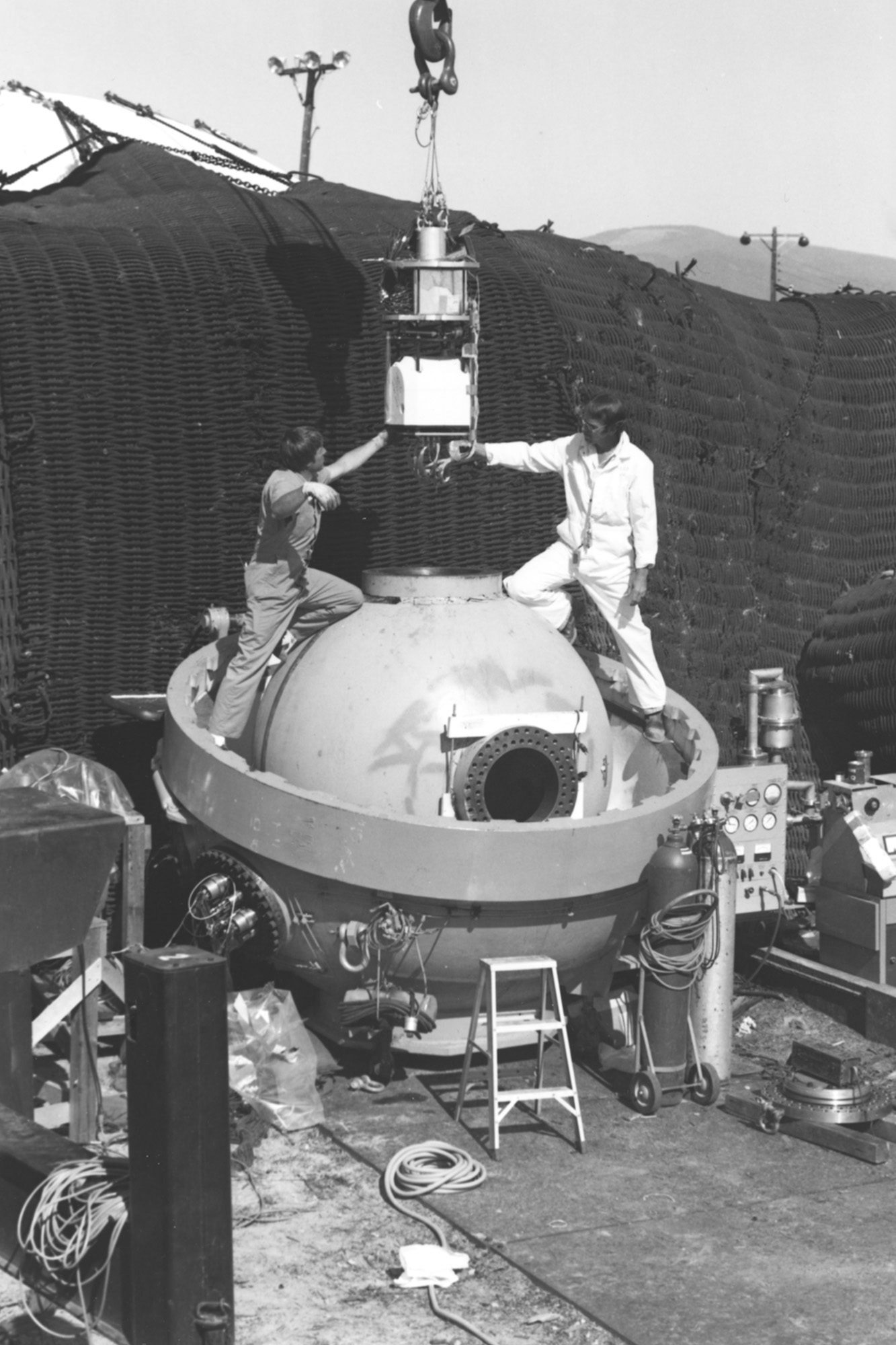
In 1961, Los Alamos scientists succeeded in generating enough high-energy photons to create flash x-ray images of explosive devices, and in 1963, the Lab opened a facility called PHERMEX: the Pulsed High-Energy Radiographic Machine Emitting X-rays. PHERMEX was the world’s first purpose-built flash radiographic facility and included many unique technological advances along with the world’s largest radio-frequency cavities. It was the first of a new generation of machines able to deliver enough energy to penetrate the metal core to create dynamic images with resolution approaching a millimeter.
“PHERMEX was the world’s first purpose-built flash radiographic facility.”
PHERMEX enabled thorough analysis of surrogate materials used to replace the fissile components in a nuclear weapon by using three large 50-megahertz radio-frequency resonators, which provided the electron beam with the energy needed for thousands of explosive experiments. Scientists used PHERMEX to confirm their design using the surrogate materials, enabling iterative tweaks. In addition, PHERMEX was used to study the fundamentals of how various materials respond in the extreme environment of a nuclear weapon—such as how they compress or expand with changes to volume, temperature, or pressure. These so-called equation-of-state data were collected for hundreds of materials, leading to an invaluable data catalog and benefitting a multitude of physics research endeavors.
PHERMEX provided vital images to the scientific community and underwent several upgrades, which increased the strength of its particle beams and changed the types of detectors to enable the highest-resolution images possible. In the 1990s, radiography became even more important for national security. For example, data on materials and implosions could be used to verify how aging weapons were expected to perform. After more than 40 years of service, PHERMEX was eventually retired, but new generations of dynamic imaging machines were developed to carry its mission forward.
A suite of capabilities
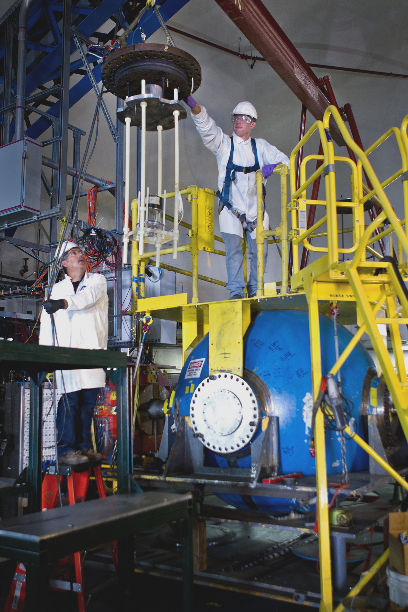
Scientists strived over the years to improve dynamic imaging tools to collect more radiographic images with improved resolution. The forces experienced in an implosion far exceed the materials’ strength and, therefore, the materials flow like liquids—prompting the experiments to be called “hydrotests.” As a result, a single, static picture often fails to capture the evolution of the implosion process.
Los Alamos scientists envisioned a new approach that would use two perpendicular beams of electrons to produce x-rays, enabling multiple pictures to be taken from two angles of an implosion inside a containment vessel. To realize this vision, the Lab built the Dual-Axis Radiographic Hydrodynamic Test (DARHT) facility, which began operation in the early 2000s and has completed over 100 hydrotests as a key part of the US dynamic imaging capability. In addition to having higher-current electron beams than PHERMEX and a smaller x-ray source spot size, DARHT incorporates a unique array of thin tubes in the image collection system, called a Bucky grid, to collimate the x-rays, improving contrast by rejecting scattered x-rays. High-speed, high-sensitivity cameras capture the unique, rapidly evolving aspects of the implosion. Images from two angles provide three-dimensional details, which cannot be identified in a single radiograph, at a higher resolution.
In addition to the DARHT facility, Los Alamos maintains an open-air firing capability called the Area 1 Firing Sites. The Firing Sites use multiple x-ray sources and detectors arranged at several angles and without the constraints found at DARHT. Scientists use this arrangement to image metal cases and their dynamic response when driven by high-explosive detonations, a technique called case radiography.
A third capability at Los Alamos uses protons instead of x-rays to create dynamic images of implosions and explosions. Taking advantage of the existing 800-million-electronvolt proton beam at the Los Alamos Neutron Science Center, Los Alamos scientists invented a new technique called proton radiography, or pRad, in 1995. The pRad facility uses the unique properties of high-energy protons to capture multiple images of a detonation. pRad employs seven solid-state, high-speed cameras that capture 21 images in a few microseconds. The result is a stop-motion radiographic movie of the explosion, giving a unique set of data for researchers not available by other methods.
Beyond the mesa
Using DARHT, the Area 1 Firing Sites, and pRad, Los Alamos scientists generate images and data that are critical to the Laboratory’s mission. Scientists at these facilities also work together with the larger dynamic imaging effort across the National Nuclear Security Administration complex, including Cygnus and the Flash X-Ray Facility.
“Using DARHT, the Area 1 Firing Sites, and pRad, Los Alamos scientists generate images and data that are critical to the Laboratory’s mission.”
Another capability, Scorpius, is a new accelerator project under construction at the Nevada National Security Sites. Scorpius will use an electron beam that can be broken into customized pulses to deliver x-rays and capture multiple images many microseconds apart.
Stemming from the earliest Manhattan Project–era attempts at visualizing implosions, Los Alamos has pioneered high-speed photography and radiography techniques that are now essential to national security. Today’s coupled capabilities ensure that our nation’s scientists have the tools they need to understand the nature of weapon components and how to safely assess the national stockpile.
People Also Ask:
- What is the difference between photography and radiography? Photography captures an image from light reflected off of an object, while radiography captures an image from high-energy particles that have passed through an object, revealing internal structures and density differences within the object.