By Levi Neukirch
Being able to see inside an object is essential to understanding how it works. X-rays revolutionized medicine by giving doctors a noninvasive way to look beneath the skin — to spot broken bones, identify problems in vital organs and detect diseases such as cancer. Now, scientists are pushing diagnostic imaging to new levels of precision with a technique called “proton radiography.” Unlike traditional X-rays, which use photons, proton radiography uses high-energy protons to create detailed images of a material’s internal structure. This approach not only provides sharper contrast but also allows researchers to observe how materials behave under extreme conditions, opening new frontiers in science.
Protons are one of the essential particles of the physical world. They are found in every atom, one of the fundamental building blocks of matter in the universe. Unlike neutrons, with which they often partner, protons carry a charge, a fact of nature that we can exploit to create images of whatever material or events the proton happens to pass through.
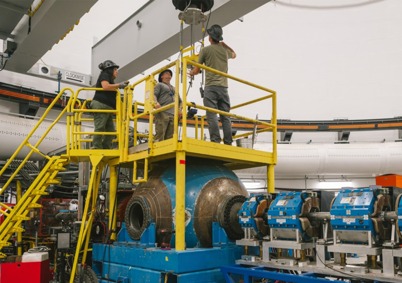
With the help of a little acceleration, the proton can be harnessed for proton radiography, creating high-resolution images of experiments in extreme and dynamic environments — a unique capability for complex but essential research. Dynamic proton radiography is a specialty made possible by the linear accelerator at the Los Alamos Neutron Science Center (LANSCE) at Los Alamos National Laboratory. At LANSCE, a beam of protons is propelled by electromagnetic fields down a half mile of pipe, reaching 84% of the speed of light.
Read the full column as it appeared in the Santa Fe New Mexican.
Contact
Media Relations | media_relations@lanl.gov