Crystal Clear
High-fidelity neutron radiography is the future of neutron science and materials characterization.
- Maya Price
- Alex Long
Meeting the nuclear demands of the nation, be it a reliable energy source or a modern nuclear stockpile, requires more than just knowledge of nuclear materials. Confidence in how these materials behave under extreme conditions or as they age is crucial.
The Los Alamos Neutron Science Center (LANSCE) plays a key role in building this confidence. As one of only five pulsed neutron sources in the world—and the only one with access to special nuclear materials—LANSCE provides a unique capability for nondestructive evaluation of materials critical to national security. Scientists at LANSCE use neutrons to probe the structure and behavior of vital materials across conditions, timescales, and environments.
Now, thanks to recent advances in detector technology, researchers at LANSCE are pushing neutron imaging forward by capturing data with higher resolution and greater fidelity than ever before. Instrument scientists Alex Long and Sven Vogel are leading this work, leveraging a state-of-the-art radiography system to streamline workflows and produce such high-fidelity images that quantitative analyses are possible. Their efforts are helping shape the future of neutron science and materials diagnostics, directly supporting the nation’s nuclear materials capabilities.
Material maps
The imaging procedure that uses x-rays is iconic: a beam of electromagnetic radiation passes through body tissue and x-rays in the beam are differentially absorbed by materials in the body. This attenuation produces a contrast pattern based on material density and the resulting image shows internal structures, like bones, with a high degree of detail.
Neutron radiography works in a similar way, but the physics behind it reveals entirely different information about the object being imaged. Whereas x-rays interact with electron clouds, neutrons interact with atomic nuclei, which means neutrons are sensitive to light elements and isotope variations. They can also penetrate dense metals that x-rays can’t, allowing researchers to see past shielding materials, fuel claddings, and other components that are opaque to other imaging techniques.
Researchers at LANSCE are pushing neutron imaging forward by capturing data with higher resolution and greater fidelity than ever before.
Because neutrons are electrically neutral, they can’t be focused or steered into a beam using magnetic or electric fields like charged particles can. At LANSCE, neutron beams are created through a process called spallation: high-speed protons from the proton accelerator are bombarded onto a tungsten target, which responds by spewing out neutrons into long, straight tubes called flight paths. For neutron radiography, the material to be imaged and a camera system are placed together within the flight path. As neutrons in the beam interact with nuclei in the sample, the neutrons are either absorbed by the material, scattered off it, or transmitted through it. As with x-rays, it’s the transmitted neutrons that create the image, but because neutrons are invisible, they must first be converted to light. The most common way of doing this is to use a neutron-sensitive material, like a scintillator, which converts the energy from incoming neutrons into photons of visible light. The light is very faint, so the camera takes long exposures, gathering enough signal over time to create a usable image.
If the neutron source can be pulsed to emit neutrons in bursts rather than one steady stream (which the LANSCE neutron source can), then the time of flight, or how long it takes each neutron to travel from source to detector, can also be measured. Because a neutron’s speed is determined by its energy, the time of flight reveals the energy of each individual neutron. The probability that an incident neutron will interact with a particular atomic species or isotope is called the neutron cross section. Cross sections are not uniform, energy-wise; neutrons of different energies have different probabilities of interacting with a given isotope, which is a key reason they make such a great materials probe. By using the measured energies of the neutrons, along with known cross section data, scientists can nondestructively study a sample material’s internal composition. The result is essentially a material map that shows which isotopes and microstructures are where across the entire sample.
An imaging system that can capture not only where within the material a given neutron went, but also when it arrived, is something that Long and Vogel are working on. They know that to capture both space- and time-resolved neutron radiography data, the camera must operate very differently from a traditional long-exposure system. First, it must be fast enough to record the exact arrival time of each neutron. But beyond speed, it must be new and fundamentally different.
A different way to take an image
Traditional neutron imaging systems are frame-based like general-use cameras: the shutter opens for a set period of time, each pixel records the total light it sees during that time, and all the pixels contribute their data to a single image, or frame.
“This is just fine for an everyday camera, but if you’re building a neutron camera to peer inside materials, this approach presents some serious challenges,” says Long.
First, traditional cameras integrate all the light detected during a fixed exposure window into a single frame. That means you can’t tell when in that interval a particular neutron event occurred—only that it happened sometime during the exposure. This is a serious limitation for time-of-flight measurements, which depend on knowing the exact arrival time of each neutron.
Second, the photons recorded by the camera don’t always come from neutrons. Gamma rays, for example, often tag along with the neutron beam or are emitted by the sample itself. These gamma rays can interact with the scintillator, and then they too are converted into visible photons and added into the mix. Even the photons that do come from neutron events can still cause trouble. When a neutron hits the scintillator, the resulting burst of light spreads out in all directions. By the time it reaches the camera sensor, what started as a small pinpoint of light appears as a larger, fuzzy glow—obscuring the precise spot the neutron hit within a given frame.
That’s why Long and Vogel are working with a new generation of neutron imaging systems, called event-mode cameras, that record light in an entirely different way. Rather than delivering time-averaged snapshots that blend together all the particles that hit the scintillator over a given time interval, the pixels of an event-mode camera operate independently from each other to record every particle interaction with the scintillator separately. The event-mode camera they use is the LumaCam, developed in recent years through a collaboration between Amsterdam Scientific Imaging and Adrian Losko, a research scientist at the Technical University of Munich in Germany (and former Los Alamos postdoc) who collaborates closely with Long and Vogel. The LumaCam delivers a data stream of time-stamped, per-pixel events, capturing the full timeline of events in a scintillator with nanosecond precision.
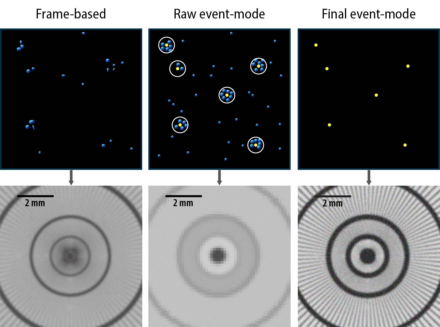
Event centroiding in frame-based vs. event-mode
What makes this approach transformative is what can be done with the data: scientists can now dissect each particle’s interaction in time and space, unlocking novel image-processing methods that are out of reach with traditional imaging to produce the highest-fidelity radiographs ever. By reconstructing the timeline of each photon burst, scientists can not only pinpoint the location of a neutron’s interaction with the scintillator, a technique known as event centroiding, but also determine the exact moment the neutron arrived. This detail is especially important for advanced neutron imaging techniques for materials science. Event centroiding is a bit like watching fireworks in the sky. Long explains, “You might not see the firework shell as it climbs through the air, but you see the pattern of light when it explodes, and from that you can figure out exactly where the shell was when it went off.”
Just as different types of fireworks can be recognized by the ways they explode—some with sharp, fast flashes, others with long, shimmering trails—different incident particles can produce subtly different light signatures on a scintillator. Scientists can use the differences in the timing and shape of each light pulse to distinguish between photons produced by neutrons and those produced by gamma rays. This forms the basis of a technique called pulse-shape discrimination, a powerful way to clean up imaging data by filtering out background photons, ensuring that only true neutron interactions make it into the final image.
With traditional imaging methods, event-centroiding is difficult and pulse-shape discrimination is next to impossible, so the images produced are qualitative. But Long and Vogel want something quantitative. Event-mode imaging moves beyond the limitations of traditional imaging by enabling new image-processing techniques. But because event-mode cameras record each pixel individually, they collect much more data than a traditional camera does—up to five gigabytes per second—and consequently, pick up much more background light. This means the raw images from an event-mode camera are actually far worse than those from traditional imaging.
All this background noise comes with an unexpected advantage: the more events the camera records, the more background noise scientists can correctly identify and remove from the data stream. After cleaning up the data through event-centroiding and pulse-shape discrimination, the images are the clearest, most accurate neutron radiographs yet, and enough to start giving quantitative answers.
A push towards quantitative neutron imaging
High-fidelity neutron images can provide meaningful physical data about a material’s composition and internal structure. To pull quantitative data from an image, scientists rely on the fundamental interactions between neutrons and matter, both at the atomic scale and across larger macroscopic features. These interactions are governed by energy-dependent neutron cross sections. The probability that a neutron with a certain energy will be absorbed by, scattered off, or transmitted through a material depends not only on the neutron’s energy and the type of atoms it encounters, but also on how those atoms are arranged in the material.
This is where LANSCE shines. Because the neutron source is pulsed, time-of-flight calculations are possible. With time-of-flight data included, it becomes possible to go beyond creating a single image, building instead a stack of energy-resolved images, each one corresponding to a specific neutron energy bin. This creates a pixel-by-pixel neutron attenuation curve, showing how a material interacts with neutrons across a range of energies at each pixel position.
Energy-resolved neutron imaging, as it’s called, can provide a wide range of information about a material—depending on the energy of the neutrons involved. The energy-dependent nature of neutron cross sections results in distinctive peaks in the attenuation curve, which indicate specific kinds of interactions. For example, at higher energies, neutrons are ideal for identifying isotopes because they interact directly with atomic nuclei. Each isotope has a unique set of absorption resonances—specific energies at which the probability of absorbing a neutron spikes dramatically. These spikes appear as sharp peaks in a material’s attenuation curve. By imaging a sample across a range of neutron energies and analyzing the shape and location of these peaks, scientists can determine the isotopes present at every pixel in the image.
“That’s the real power of neutron radiography at this level. It moves beyond showing where things are to reveal what they’re made of and how they’re structured.”
At lower energies, neutrons interact more strongly with the atomic lattice. In crystalline materials, this can lead to a change in the likelihood of scattering versus transmission at specific energies. These changes appear in attenuation curves as a series of step-like features known as Bragg edges (named for Lawrence Bragg and the characteristic shape of the curve). Each Bragg edge corresponds to a particular set of crystallographic planes and provides information about the material’s crystal structure, phase distribution, and grain orientation. Taken together, absorption resonances and Bragg edges make it possible to probe materials from the nuclear scale to the atomic scale using a single imaging technique.
When isotopic composition and crystalline structure are resolved at the pixel level, a neutron image becomes more than just a visual—it becomes a truly quantitative material map. But achieving that level of precision requires exceptionally clean data. The subtle features in a material’s attenuation curve—like Bragg edges and absorption resonances—can be obscured by background signals and noise. That’s why Long and Vogel are pursuing event-mode imaging. By recording each neutron interaction individually, with precise timing and spatial resolution, event-mode systems dramatically reduce background and make it possible to extract meaningful, quantitative results.
“That’s the real power of neutron radiography at this level,” says Vogel. “It moves beyond showing where things are to reveal what they’re made of and how they’re structured.”
Vogel and Long have taken neutron imaging at LANSCE from a simple imaging technique to an advanced tool for materials characterization. Their quantitative neutron imaging method is already being applied to a range of materials important to national security and nuclear energy, from irradiated nuclear fuels to structural materials. The method can proactively confirm a new material’s microstructure to ensure it will perform as expected, but it can also retroactively reveal the effects of various extreme conditions and thereby guide the design of next-generation materials and nuclear fuels.
No neutrons left behind
Imaging can reveal a great deal about a material, but it is not the most sensitive probe. That’s because imaging relies on detecting small changes in a large quantity—the overall beam intensity—and the resulting signal is spread out across many pixels. By contrast, diffracted neutrons, which scatter in many directions rather than passing straight through a material, can provide complementary information with far greater sensitivity to subtle structural details.
“Materials are like obstacle courses for neutrons,” explains Long. “Some neutrons make it straight through to the detector, giving us the transmitted signal that forms an image. Others scatter or diffract along the way. Those ‘detoured’ neutrons carry information that is far more sensitive to the material’s underlying structure than the mere absence of transmitted neutrons in an image.”
Diffraction with pulsed sources also relies on time-of-flight techniques: because a neutron’s wavelength must correspond to the spacing of the crystal lattice, its energy influences if and how it scatters. Indeed, diffraction is especially sensitive to microstructural properties and can reveal additional details of the crystalline structure—like phase distribution or grain orientation changes. The tradeoff is that diffraction data are averaged over the entire neutron-illuminated volume, so while the sensitivity is high, the results lack spatial resolution. Diffraction data alone can’t show the location or spatial distribution of the microstructural features it detects, only that they are present and in what proportion. That’s why the combination of imaging with diffraction is so powerful. Precise diffraction data can be used to guide and verify interpretation of a neutron image, and the image in turn can be used to map the spatial distribution of the quantities measured by diffraction. Capturing both sets of neutrons—the ones that pass through and the ones that diffract—leads to a more complete understanding of a material. The final product is a detailed map showing the nature, location, and quantity of every variation in the material’s composition, across both micro- and macroscopic scales.
At LANSCE, imaging and diffraction have traditionally been performed separately. This burns time and resources: twice the beam supply, separate instrument setups, and careful coordination to ensure that conditions are replicated across experiments. It also complicates data analysis, because the datasets must be reconciled. But all this is changing with the arrival of the LumaCam event-mode camera that Long and Vogel have been using. Its flexible, modular design makes it easy to integrate with existing technologies. Long and Vogel have also now proven that LumaCam makes it possible to collect both imaging and diffraction data from the same experiment simultaneously.
This approach makes it possible to collect both imaging and diffraction data from the same experiment simultaneously.
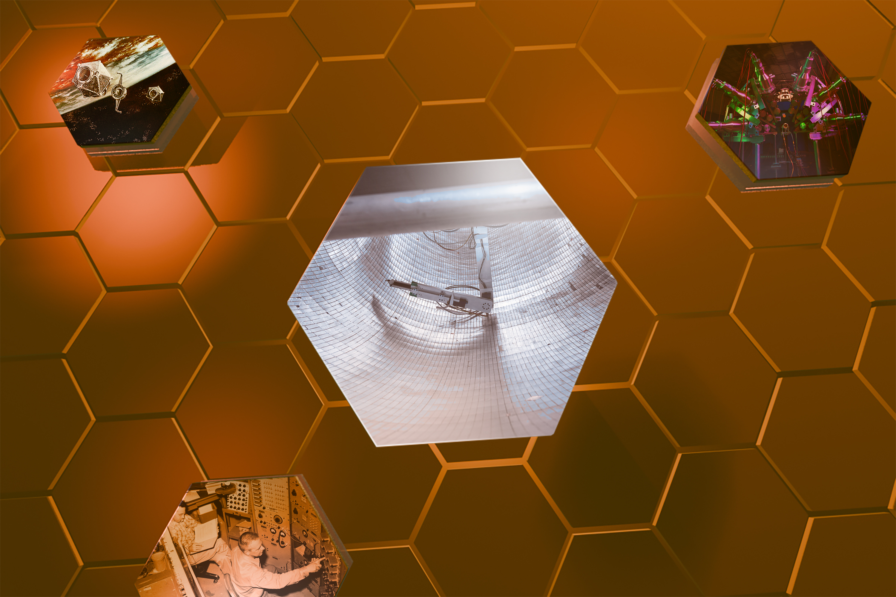
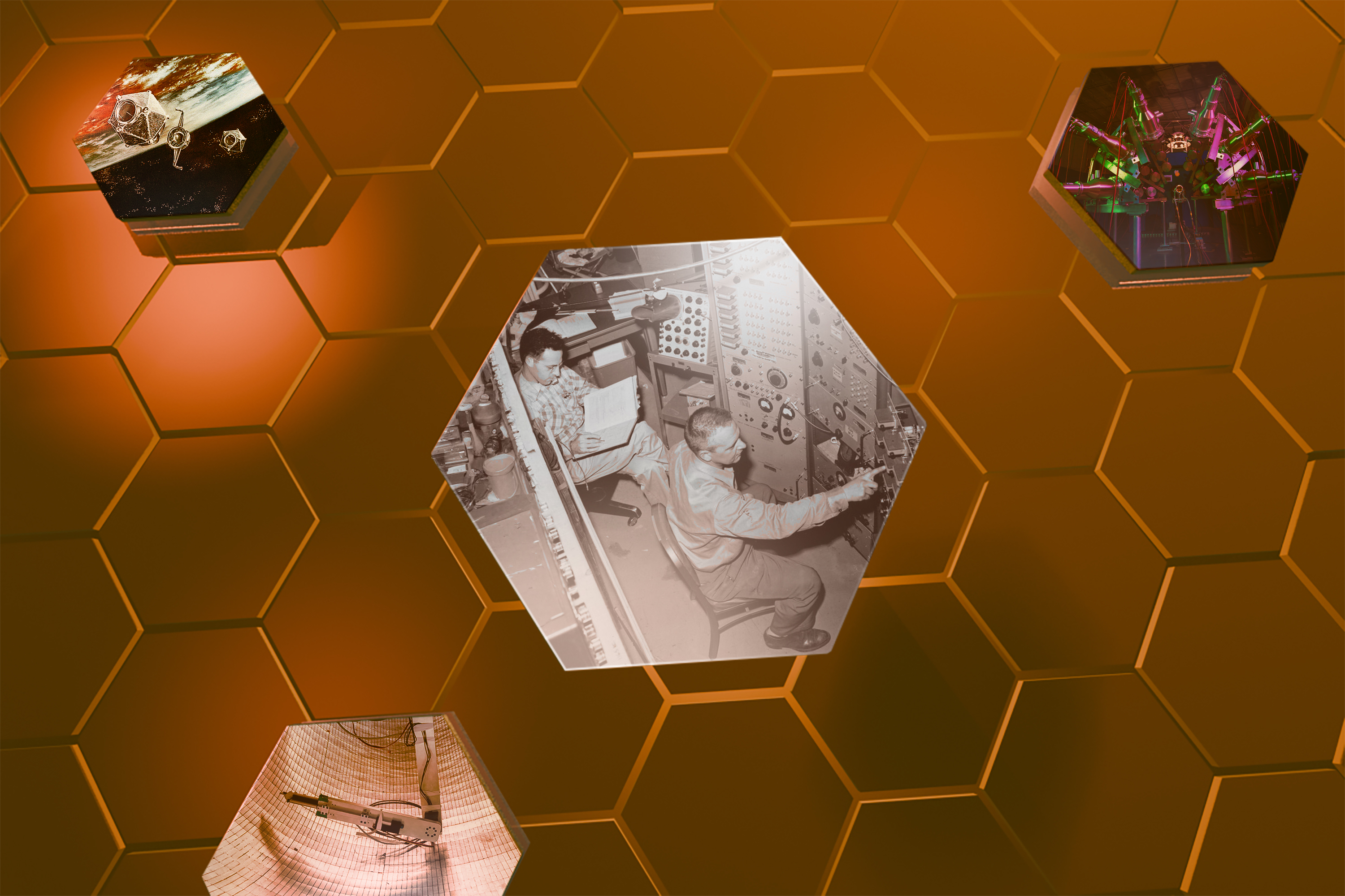
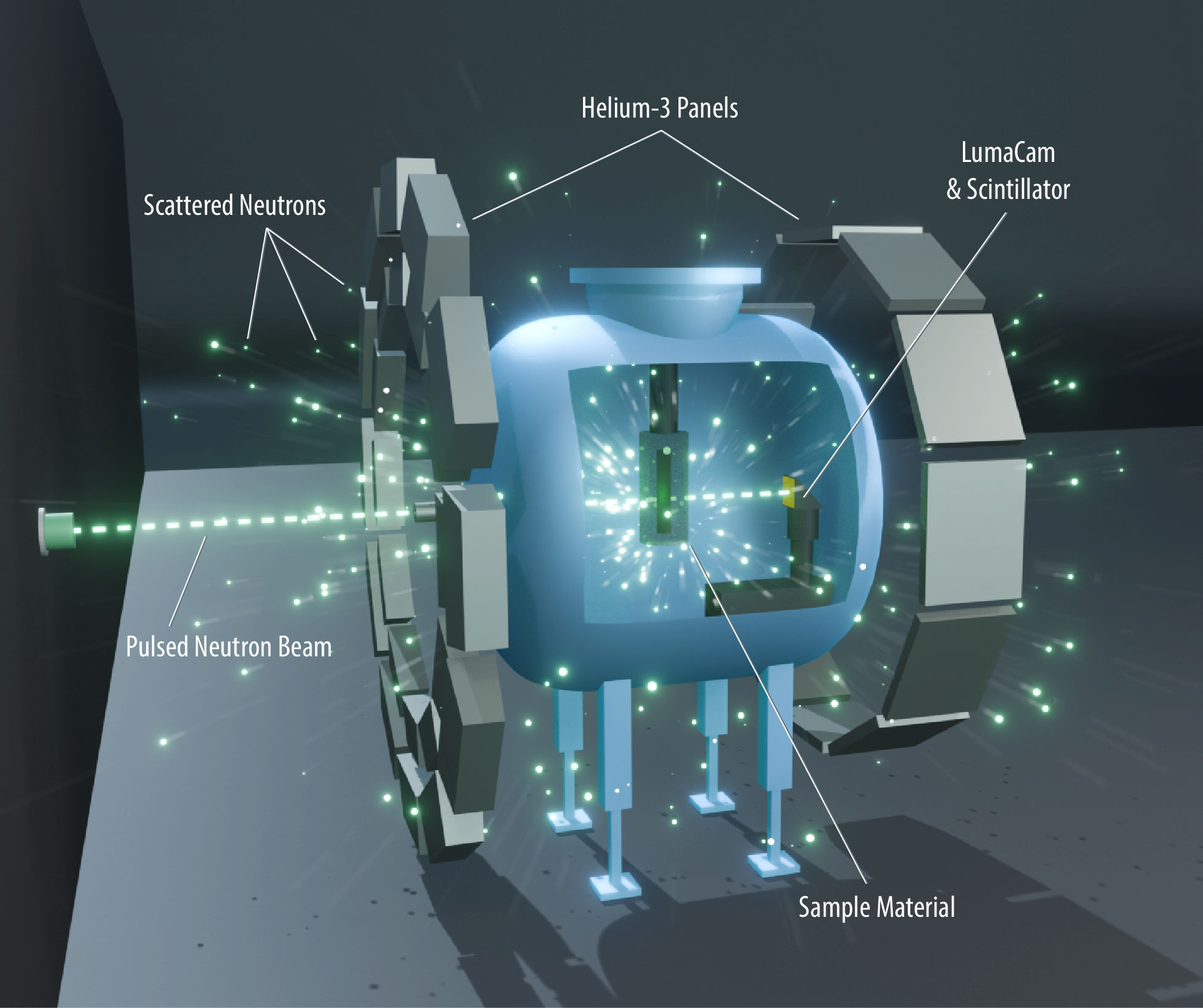
As a proof of principal, the scientists retrofitted a LumaCam on the High-Pressure Preferred Orientation (HIPPO) diffractometer at LANSCE. HIPPO is a time-of-flight neutron diffractometer equipped with 1,200 detectors, each one a thin metal tube filled with helium-3 (³He) gas, positioned all around the sample. By placing the LumaCam just downstream of the sample, they were able to capture both transmitted and scattered neutrons at the same time—without moving the sample or switching instruments. This dual-detection setup enables high resolution, spatially resolved images and sensitive bulk diffraction measurements in a single experiment.
The team has already demonstrated the effectiveness of this combined technique across a range of complex materials. Because it’s better in just about every way—faster and more beam efficient because less repetition is needed, cheaper because costly custom electronics aren’t required, and more reliable because the two data sets are more readily integrated thereby reducing uncertainty—Long and Vogel are eager to see it implemented across more neutron flight paths at LANSCE.
Looking forward
Now that they have a way to detect the time and location of every neutron’s arrival, the team is exploring other areas of neutron science. They want to show the utility of the LumaCam as not just as an imaging tool, but as a full replacement for traditional neutron detectors in various other settings.
The 1,200 detectors on the HIPPO diffractometer are a perfect example. Each ³He-filled tube requires high voltage power and custom-built electronics, not to mention ³He itself is increasingly hard to source. Legacy systems like these are expensive and impractical for operations and maintenance and when it comes to building new beamlines. The LumaCam is a promising alternative: it’s compact, commercially available, comparatively low-cost, and requires no special supporting hardware.
Beyond imaging and diffraction, the team has also demonstrated the LumaCam’s potential use in other kinds of neutron experiments, including reflectometry and small-angle scattering. No matter what it’s being used for, the core hardware remains the same—the only thing that changes is how the data are processed. Although processing data from a LumaCam does require more computing resources, those resources are already in place and the tradeoff brings enormous flexibility and potential.
This kind of modularity simplifies everything. As neutron science becomes increasingly important to the Laboratory’s ability to meet the nation’s energy and national security needs, so too will multi-modal material characterization. Thanks to flexible new technologies and innovative thinking, scientists are already spending less time managing instruments and more time pushing the boundaries of science.
People also ask:
- What is neutron imaging? Neutron imaging is a form of nondestructive testing that uses neutrons to probe materials, creating images of a material’s internal structure and features. It’s a similar concept to x-ray imaging, but because neutrons interact with matter in different ways than x-rays, they can “see” certain things that x-rays and other imaging probes cannot.
- What is materials science? Materials science studies the relationships between a material’s properties, structure, and behavior, drawing from multiple areas of science and engineering. Understanding these relationships is the basis for improving existing materials and developing new ones.