The Far-reaching Legacy of Flow Cytometry at Los Alamos National Laboratory
In 1965, a Los Alamos scientist named Mack Fulwyler was evaluating blood cells in solution and needed a way to quickly identify and isolate specific types of cells for further study. After reading about the invention of the ink-jet printer, which uses vibration to create a stream of individual tiny ink droplets, he was inspired to create something new. Fulwyler’s prototype flow cytometer suspended blood cells in droplets of solution, making them easier to separate and sort than if they were in a steady stream. Fulwyler’s work, combined with other inventions at Stanford University and the University of Münster in Germany, led to the development of modern-day flow cytometers and cell sorters which are now ubiquitous in medical and research laboratories worldwide.
How it works:
The basic principle of flow cytometry is that cells or particles, suspended in a liquid stream, flow single-file past a set of lasers, enabling detection and sorting based on light scatter and/or fluorescence. This process allows for multiplexed measurements on individual particles, with a large number being detected in a short period of time. Additionally, selected populations of those particles can be isolated for further analysis, or regrowth in the case of viable biological particles.
National Flow Cytometry Resource 1982-2013
For over three decades Los Alamos was home to the National Flow Cytometry and Sorting Research Resource (NFCR),which was funded by the National Institutes of Health. The NFCR funding supported the development of new flow cytometry instrumentation and research techniques, as well as a hands-on Annual Course in Flow Cytometry, that was held on alternate years in either Los Alamos, New Mexico or Brunswick, Maine, and continues at other institutions today.
Schematic for the early fluorescence activated cell sorter (circa 1965) invented by Los Alamos scientist Mack Fulwyler. Flow cytometers are now used worldwide in research and clinical labs.
Sorting Technologies and the Human Genome Project
The early years of the NFCR was a time for rapid development of instrumentation and new applications. One of the first areas of R&D was focused on identification of chromosome alterations related to genetic disease or DNA damage. Methods for chromosome sample preparation were developed, and chromosome analysis and sorting techniques were refined by a combination of improved chromosome staining methods and instrumentation development.
Building on these capabilities, researchers then had the idea of making DNA libraries for all 24 human chromosome types, for use in mapping disease genes to specific chromosomes. This idea was the basis for the DOE National Laboratory Gene Library Project (in collaboration with Lawrence Livermore National Laboratory), in which researchers used flow cytometry to create libraries of human chromosomes for distribution to researchers worldwide. This capability was key to the origins of the Human Genome Project by ensuring that enough copies of DNA would be available for sequencing.
In parallel, during these early years, NFCR researchers were developing improved methods to stain specific cell components: DNA, protein, RNA-and using these fluorescent dye combinations to study multiple components of each cell during the cell cycle, or in relation to a specific cancer cell type. They developed one of the first multiparameter flow cytometers with 4 different lasers and multiple detectors to differentiate the polychromatic dyes, each staining a different cell component.
Throughout the term of the NFCR many new instrument technologies and applications were developed, some of these are described below:
Ultrasensitive Flow Cytometry
Ultrasensitive Nucleic Acid Detection: Los Alamos scientists used fluorescence correlation spectroscopy of single molecules combined with novel labeling strategies to detect nucleic acid targets at the zeptomole level. This approach was developed for RNA detection and molecular haplotyping.
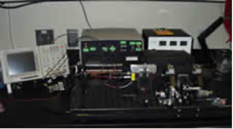
High Sensitivity Flow Cytometer: This was one of the first flow cytometers able to push the lower limit of detection to smaller than a bacteria. Using very low excitation power (typically < 2 mW @ 532 nm) and high sensitivity fluorescence detectors (including photon-counting APDs) it could detect very small particles (100 nm dia. beads) or very weakly fluorescent particles. This was due primarily to its slow flow velocity (typically 1 cm/sec). This instrument was used primarily to measure the length of individual DNA fragments; the resulting histogram showed series of peaks, similar to what you would see on a gel running restriction fragments. This method required much less DNA, and took only minutes to run a sample. Other small particle applications included individual mitochondria and intact viral particles.
High Sensitivity Flow Cytometer
Bead-based microarray/SNPs approach: Single-nucleotide polymorphisms (SNPs) are the most abundant type of human genetic variation. These variable sites are present at high density in the genome, making them powerful tools for mapping and diagnosing disease-related DNA sequence variations. Los Alamos scientists developed a sensitive and rapid flow cytometry-based assay for the multiplexed analysis of SNPs based on polymerase-mediated primer extension, or minisequencing. In the assay, biotinylated primers were captured onto streptavidin-coated microspheres, and the number of incorporated fluorescent ddNTPs was measured by flow cytometry.
Bacterial Bioforensics: As part of a consortium of National Laboratories, Los Alamos scientists applied state-of-the-art molecular analysis methods to the forensic analysis of bacterial pathogens. The DNA Fragment Sizing Flow Cytometer enabled restriction fragment analysis of whole genomes to fingerprint organisms without the need for DNA sequence information. For analysis of DNA sequence-based signatures, we used SNP analysis on microsphere arrays to interrogate functionally- and phylogenetically important sequences.
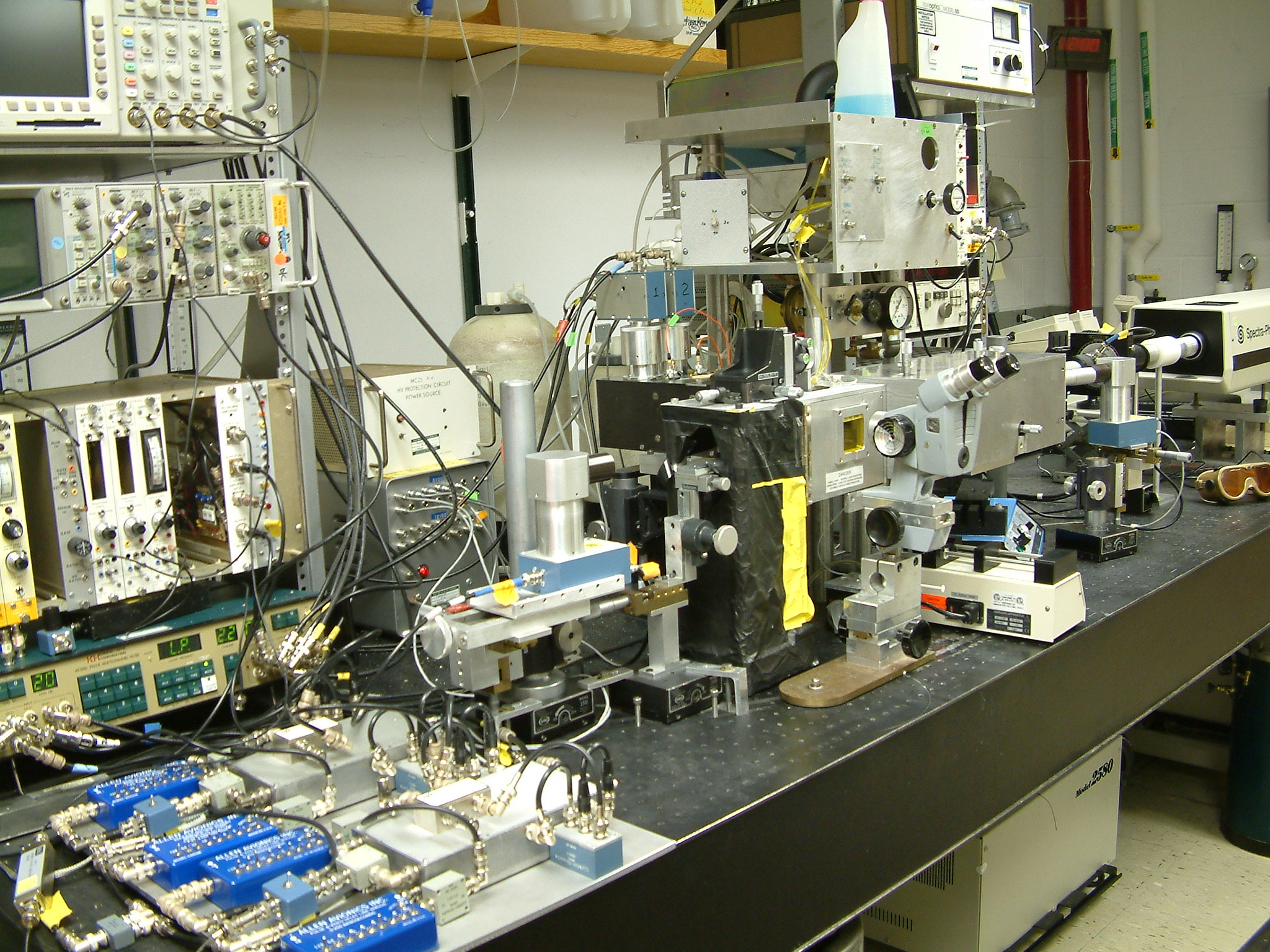
Spectral and Fluorescence Lifetime Flow Cytometers
Several types of spectral flow cytometers were developed at Los Alamos with the aim to capture and interpret the information contained in the excitation and emission spectra of fluorescent compounds whether naturally occurring from cell constituents or applied to the cells in the form of a fluorescent dye.
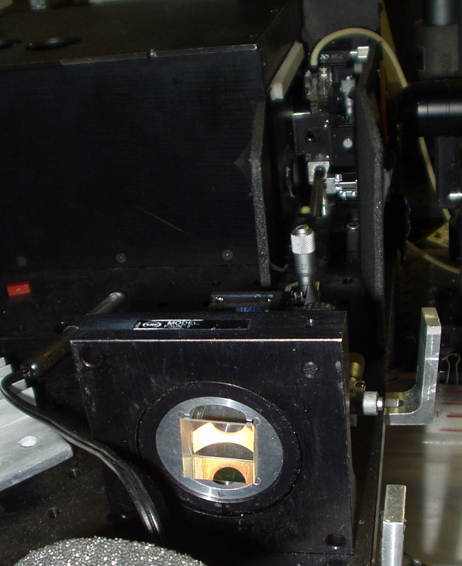
Fourier-transform Flow Cytometer: The first instrument developed in the NFCR to measure fluorescent spectra in cells was the Fourier-transform flow cytometer. The Fourier-transform flow cytometer used a high-speed interferometric approach to determine the intensity spectra of individual cells in a flow system. The Fourier-transform flow cytometer employed photomultiplier tubes as detectors, which enabled the rapid measurement of many cells, but interrogating only one emission band at a time.
Fluorescence Lifetime Flow Cytometer: Detection of dyes with overlapping spectra are a challenge in traditional flow cytometry, leading to complicated compensation matrices or inability to use certain dyes. The fluorescence lifetime flow cytometer took advantage of another property of dyes, their fluorescence lifetime, which is the time between dye excitation and emission of fluorescence. By using a modulated laser beam the phase shift of the light emitted by a fluorescent dye could be detected and the fluorescence lifetime calculated. If two different dyes had overlapping spectra but different lifetimes they could be easily separated. The instrument was also used to directly measure the fluorescent lifetime and to separate fluorescence of a label from autofluorescence or light scatter.
Fluorescence Lifetime Flow Cytometer
Spectral Analysis Cytometer: The NFCR full-spectral analysis flow cytometer had a wavelength resolution of ~2 nm over the range of 350-800 nm and a single particle intensity
sensitivity of ~150 MESF of FITC. This analytical instrument was designed for a more comprehensive and flexible approach to spectral analysis including: spectral deconvolution of overlapping emission spectra, fluorescence resonance energy transfer measurements, metachromic dye analysis, free versus bound dye resolution, and Raman spectroscopy.
Spectral Analysis Cytometer
Acoustic Flow Cytometry
Although hydrodynamic focusing used in traditional flow cytometers is a very effective approach to position particles for analysis, the use of sheath increases assay cost and reduces instrument utility for field and autonomous remote operations. The acoustic flow cytometer was developed to use acoustic excitation, generated along the entire structure of a capillary tube, to both focus and concentrate sample particles to the interrogation region. Because acoustic methods both focus and concentrate particles, it is possible to maintain both conventional particle analysis rates as well as long transit times while using a fraction of the power and consumables of a conventional flow cytometer. The longer integration time allows conventional particle analysis using data acquisition systems that are less expensive, smaller and require less power, while still performing high sensitivity measurements. However, the benefits of acoustic focusing flow cytometry are not restricted to only the elimination of sheath fluid. Acoustic concentration also enables the analysis of extremely dilute samples on the order of several cells or particles per liter, as might be seen in a water monitoring application, at reasonable analysis rates. Since there is no sheath, it is also possible to repeatedly reanalyze particles of interest for reliable rare event analysis.
Acoustic Flow Cytometer
In addition to winning several awards, the acoustic flow cytometer was successfully commercialized as a start-up company called Acoustic Cytometry Systems, which was later bought by Invitrogen. In 2008, ACS was acquired by Invitrogen, was purchased by Life Technologies, and subsequently Thermo Fisher and is now marketed as the Attune Flow Cytometer.
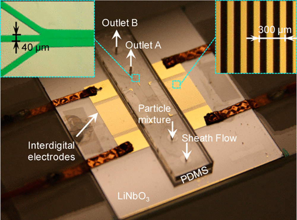
In the final years of the NFCR, we successful applied standing surface acoustic waves to the separation of Escherichia coli bacteria from peripheral blood mononuclear cells, in a microfluidic, flow sorting “lab-on-a-chip” device and demonstrated efficient collection of the separated cells.
Photograph of the fabricated microfluidic device for particle separation. The upper-left inset is the zoomed-in view of the three-outlet junction. The upper-right inset is the zoomed-in view of the interdigital transducers.
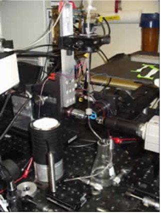
Rapid Mix Flow Cytometry
Molecular interactions occur at a high rate of speed and in flow cytometry, the earliest analysis point is limited to the speed at which the sample can be introduced into the flow cell. The Rapid Mix Flow Cytometer had an injection point that allowed for the mixing of one reagent to another within the sample line directly before the flow cell. The injection point was variable, which allowed the user to vary the amount of time a sample was mixed prior to analysis, the shortest being 120 ms.
Rapid Mix Flow Cytometer
Data Analysis for Flow Cytometry
Beginning in the 1970’s, and through the years that the NFCR existed, data analysis methods were needed to support the different types of data being acquired from new flow cytometry and sorting instrumentation and their applications. Researchers at Los Alamos developed new data analysis tools, as well as new data acquisition technologies. In the NFCR, in work that began in the 1990’s. we designed, fabricated, and implemented a new general system for flow cytometric data acquisition and analysis. This system called the Digital Data Acquisition and Control system—DiDAC—was developed to acquire data, control sorting, and have a computer-based user interface for control of numerous instrument parameters and to provide new data display and analysis capabilities. DiDAC was initially developed to support data acquisition capabilities for unique NFCR research instruments and for collaborators, but was envisioned to have a large impact on the broader flow cytometry community. The DiDAC technology was matured and updated to the Open Reconfigurable Cytometric Acquisition system (ORCAs), to support novel flow cytometry efforts, and later in the final years of the NFCR, was spun off to a Los Alamos small business for commercialization.

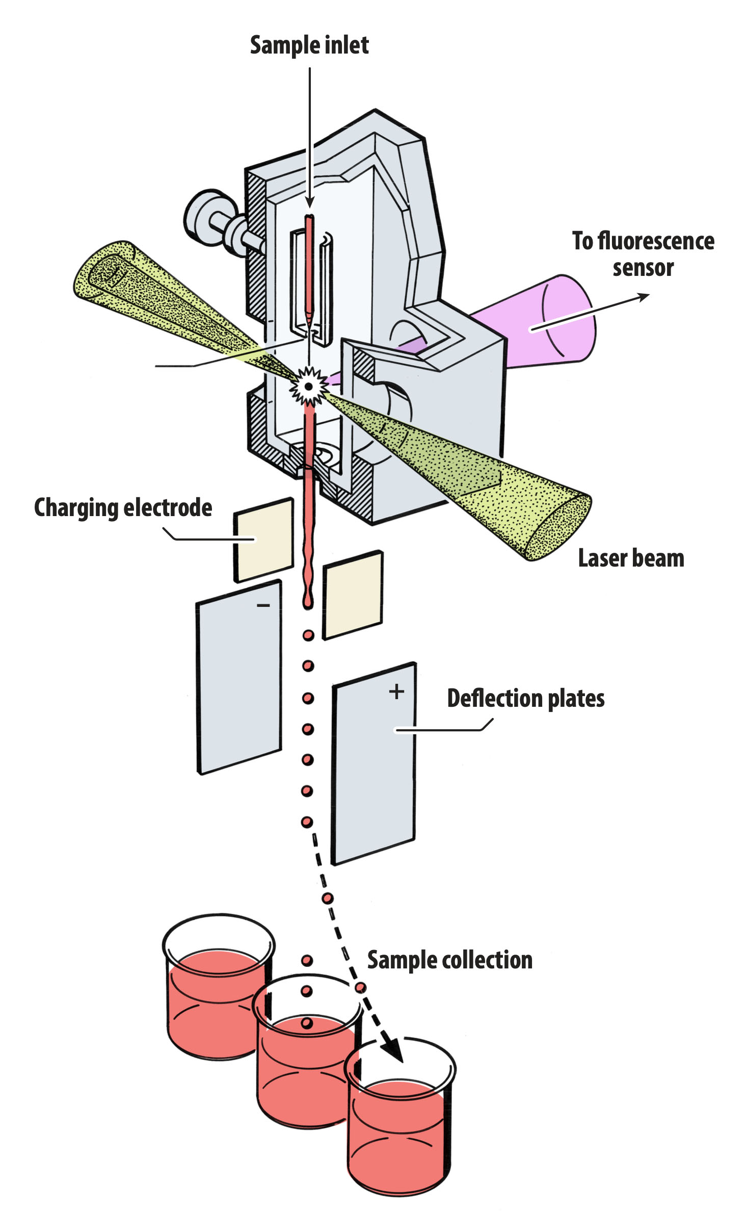 Schematic for the early fluorescence activated cell sorter (circa 1965) invented by Los Alamos scientist Mack Fulwyler. Flow cytometers are now used worldwide in research and clinical labs.