PHYSICAL vs MATHEMATICAL ANTHROPOMORPHIC
PHANTOMS:
A COMPARISON BETWEEN REAL AND PREDICTED
ACTIVITIES
1Bernardo
M Dantas, 1John G Hunt, 2Henry B Spitz & 3Irena
Malátová
1Instituto
de Radioproteção e Dosimetria
Av
Salvador Allende - s/n - Rio de Janeiro - RJ - Brazil - 22780-160
2University of Cincinnati
598 Rhodes Hall - Cincinnati
- Ohio - USA - 45221-0072
3National
Radiation Protection Institute
Srobarova
48 - 10000 Prague 10 - Czech Republic
ABSTRACT
The
Instituto de Radioproteção e Dosimetria (IRD-CNEN) Whole Body Counting Facility
in Brazil uses an array of four high resolution germanium detectors for routine
in vivo measurements of 238U
(Th-234), 235U, 241Am and 226Ra. The system is able to detect radionuclides
emiting photons in the energy range
from 10 to 200 keV. Detectors are calibrated using conventional physical
anthropomorphic phantoms including the Livermore thorax phantom for
radioactivity deposited in the lungs and knee and skull phantoms for
radioactivity deposited in bone.
Recently, mathematical phantoms using Monte Carlo analysis have been
used to determine calibration factors for in
vivo measurements. Monte Carlo
analysis is used to simulate the interactions associated with the transport of
photons from an origin in human tissue to the detector for a unique counting
geometry. The computer code currently in use at IRD consists of a routine
developed using Visual Basic specifically for this application. The code,
Visual MC, simulates photon transport within a voxel phantom and the detection
of those photons that escape and enter the detector. The phantom used was
provided by the National Radiological Protection Board (NRPB) and contains 3.6
x 107 voxels,
each one measuring 0.208mm x 0.208mm x 0.202mm. The accuracy of Visual MC was verified
through the comparison of benchmark measurements of phantoms with the predicted
activity. Results for 226Ra
and 241Am in the knee, 241Am and 152Eu in the
skull and 241Am, 238U and 235U in the lungs
show that the predictions agree favorably with the benchmark measurements and
produce an uncertainty comparable to that obtained during a typical in vivo measurement.
INTRODUCTION
The
direct measurement of radionuclides in the human body has become one of the
most useful tools in the field of radiation protection and dosimetry. In vivo
measurements of radionuclides emitting low energy photons are usually performed
in heavily shielded rooms equipped with highly sensitive detection systems in
order to attain the dose limits established by regulatory organizations (IAEA,
1996; ICRP, 1997). The calibration of such devices is routinely carried out
using physical anthropomorphic phantoms containing well-known amounts of the
radionuclides of interest (ICRU, 1992). On the other hand, a number of
facilities have recently dedicated efforts for the development of mathematical
phantoms and computer codes in an attempt to simulate the physical phenomena of
interaction of radiation with matter using voxel phantoms and the Monte Carlo
method (Breismeister,
1993; Dimbylow, 1996; Mallett et al,
1995). The application of this
technique to the calibration of in vivo detection systems is likely to
become a worthy alternative in the cases where no conventional phantoms are
available. The possible applications include the quantification of wound
contamination (Hickman et al, 1994). It can also be a useful method in the
planning and design of new physical phantoms and detectors (Kramer et al,
2000a) as well as for the optimization of counting geometries and the
estimation of errors in parameters involved in direct measurements, such as
detector positioning, radionuclide distribution within the organ or tissue and
organ size (Kramer and Crowley,
2000; Kramer et al, 2000b; Kramer et al, 1997; Kramer and Yiu, 1997). This method is not intended to replace
the conventional phantoms for whole body counting calibration, but to
complement them with a quick, versatile and low cost procedure.
MATERIALS AND METHODS
The
IRD-CNEN Whole Body Counter uses an array of four HPGe detectors for in vivo measurements of
radionuclides emitting photons in the energy range from 10 to 200 keV. The
detectors are 20-mm thick, with an active diameter of 50.5 mm, active areas of
2000 mm2 and 0.6 mm carbon composite window. Each pair of detectors
is connected to a 3-liter cryostat. The resolution (FWHM) specified for this
system is 370 eV at 5.9 keV and 675 eV at 122 keV. Detectors are set to present
the same energy calibration. The curve can be expressed as E(keV) = 11.29 +
0.2096 x Channel Number.
The
measurements are performed inside a 2.5m x
2.5m x 2.62m room with 15 cm steel walls, each covered internally with a 3 mm lead
layer, followed by a 1.5 mm cadmium layer
and finally a 0.5 mm copper layer. These layers reduce the background count
rate mainly in the energy range below 200 keV (Oliveira at al, 1989).
With
the objective of providing a wider range of calibrations for specific internal
contamination circumstances and counting geometries the use of mathematical
phantoms for whole body counting calibration has been tested and a computer
code called Visual MC has been developed (Hunt, 1999). The mathematical phantom
used was supplied by NRPB and contains 3.6 x 10⁷
voxels. In order to verify the suitability of the mathematical phantom chosen
to simulate the organs and tissues of interest and the accuracy of the computer
code developed specifically for this application, a series of comparisons
between calculated and real activities contained in some selected physical
phantoms has been performed. The physical phantoms used in this comparison
represent the most usual calibration techniques for the measurements of low
energy photon emitters monitored routinely.
The
conventional phantoms used for benchmarking include the LLNL (Lawrence
Livermore National Laboratory-CA-USA) thorax and lungs (Griffith et
al, 1984), UC (University of Cincinnati-OH-USA) lungs, knee and skull (Spitz,
2000), the NRPI (National Radiological Protection Institute-CZR) skull phantom
(Malatova
and Foltanová , 2000; Malatova and Foltanová,
1988; Ruehm
et al, 1988), the BfS skull and the USTR case 102 skull.
In
order to accumulate a significant number of counts in each specific region of
interest, phantoms and point sources of each radionuclide of interest were
counted inside the shielded room with different count times and distances to
the detector, depending on their activities. The counting parameters are shown
in Table 1.
The Monte Carlo technique is used to simulate a tissue contamination, to
transport the photons through the tissues and to simulate the detection of the
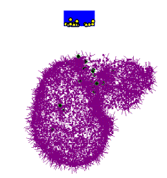
radiation. The graphic output of Visual MC shows the counting geometry and the
photon interactions., see Figure 1.
A voxel phantom with a format of 871 “slices” each of 277 x 148 picture
elements was used. The voxel phantom is derived from a whole body Magnetic
Resonance Image (MRI) scan with contiguous slices (Dimbylow, 1996). The scanning data were kindly supplied by the
Non-Ionising Radiation Department of the National Radiological Protection Board
(NRPB). The size of each voxel is 2.08 mm x 2.08 mm x 2.02 mm. The voxel
phantom was scaled so that the height (1.76 m) and the mass (73 kg) conforms to
the values for reference man stated in ICRP 66 [10]. The phantom is called
NORMAN, which stands for Normalised Man. There are 37 tissue types represented
in the NRPB version of NORMAN. The version of NORMAN supplied by NRPB for this
work is divided into the following tissue types: adipose tissue, hard bone,
bone marrow, thyroid and lungs. The
other tissues and muscles are grouped together.
Table
1. Measurements of phantoms and point sources
|
Phantom |
Phantom
data |
Point
source data |
||||||
|
Dist (cm) |
Area (counts) |
CT
(min) |
Activ
(Bq) |
Dist
(cm) |
Area (counts) |
CT
(min) |
Activ
(Bq) |
|
|
aAm-241-Lungs |
7.5 |
f39461 |
60 |
18577 |
50 |
25565 |
10 |
199166 |
|
aAm-241-Skull |
9.0 |
g126696 |
60 |
58802 |
50 |
25565 |
10 |
199166 |
|
aAm-241-Skull |
14.5 |
h87207 |
60 |
58802 |
50 |
25565 |
10 |
199166 |
|
aAm-241-Tibia+Fibula |
8.0 |
9822 |
10 |
13062 |
50 |
25565 |
10 |
199166 |
|
bAm-241-Skull |
3.0 |
1.27 x 10⁷ |
60 |
700000 |
5.5 |
28668 |
10 |
17720 |
|
cAm-241-Skull |
3.0 |
4.81 x 10⁵ |
666 |
5400 |
5.5 |
28668 |
10 |
17720 |
|
dAm-241-Skull |
3.0 |
1062 |
30 |
620 |
5.5 |
28668 |
10 |
17720 |
|
aRa-226-Lungs |
7.5 |
f831 |
1000 |
1859 |
50 |
2335 |
10 |
328971 |
|
aRa-226-Tibia |
7.5 |
3505 |
1000 |
441 |
50 |
2335 |
10 |
328971 |
|
aEu-152-Skull |
9.0 |
g40705 |
60 |
20434 |
40 |
4417 |
10 |
31993 |
|
aEu-152-Skull |
14.5 |
h27601 |
60 |
20434 |
40 |
4417 |
10 |
31993 |
|
eU-235-Lungs |
2.94 |
f1057 |
60 |
394 |
6.5 |
31484 |
120 |
10.007 |
|
eU-238-Lungs |
2.94 |
f1824 |
60 |
8451 |
6.5 |
704 |
60 |
214.6 |
|
eU-238-Lungs |
2.94 |
f2563 |
60 |
8451 |
6.5 |
1067 |
60 |
214.6 |
a. University of
Cincinnati-OH-USA; b. National Radiological Protection Institute-CZR; c.
BfS-Germany; d. BPAM-01-USTUR case 102; e. Lawrence Livermore National
laboratory-CA-USA;
f. Center chest; g. Front
head; h. Right side
Before making the calculation with the NORMAN phantom, it is
necessary to “calibrate” the mathematical detector by counting a point source
of the same radionuclide on the center line of the detector. This “calibration”
allows the real detector losses in the window and electronics to be compensated
for during the calculation.
RESULTS AND CONCLUSIONS
The
counting geometries were simulated mathematically and the activities contained
in each phantom were calculated using the mathematical phantom and the
Visual-MC code. Results of the comparison between real and calculated
activities are shown in Table 2. The results show that the calculated
measurements agree favorably with the real activities. It is important to point
out that the accuracy of this type of mathematical simulation depends on the
similarity between the counting geometry of the physical phantom and the
mathematical phantom. Although the improvement of certain features of the code
are still under development, its output already produces an uncertainty
comparable to that obtained during a typical in vivo measurement.
Table
2. Comparison of phantoms activities with predictions made using Visual-MC code
|
Radionuclide |
Geometry |
Activity
(Bq) |
%
error |
||||
|
Real |
Calculated |
||||||
|
Am-241 |
Lungs
(center chest) |
18577 |
18900 |
+
1.74 |
|||
|
Am-241 |
Skull
(Frontal) |
58802 |
47900f |
-
18.5 |
|||
|
Am-241 |
Skull
(Lateral) |
58802 |
51900l |
-
11.7 |
|||
|
Am-241 |
Tibia+Fibula |
13062 |
12300 |
-
5.83 |
|||
|
Am-241 |
Skull
(Lateral) |
700 |
970 |
+
38.6 |
|||
|
Am-241 |
Skull
(Lateral) |
5400 |
6700 |
+
24.1 |
|||
|
Am-241 |
Skull
(Lateral) |
620 |
480 |
-
22.6 |
|||
|
Ra-226 |
Lungs
(Frontal) |
1859 |
2130 |
+
14.6 |
|||
|
Ra-226 |
Tibia |
441 |
505 |
+
14.5 |
|||
|
Eu-152 |
Skull
(Frontal) |
20434 |
20600f |
+
0.81 |
|||
|
Eu-152 |
Skull
(Lateral) |
20434 |
19600l |
-
4.08 |
|||
|
U-235 |
Lungs
(Center chest) |
394 |
471 |
+
19.5 |
|||
|
|
Lungs
(Center chest) |
8451 |
7692 |
-
8.98 |
|||
Figure 1: Counting geometry for
the head phantom, 241Am, front head, bone surface. The circles in the
germanium detector represent photoelectric interactions.
REFERENCES
BREISMEISTER, J.F. MCNP - A
general Monte Carlo code for N-particle transport code version 4A. Los Alamos,
NM: Los Alamos National Laboratory; LA-12625-M (UC705 and UC700), Rev. 2; 1993.
DIMBYLOW P.J. The development of realistic voxel phantoms for
electromagnetic field dosimetry. In: Voxel Phantom Development, Proceedings of
an International Workshop, National Radiological Protection Board, Chilton,
1-7, 1996.
GRIFFITH, R.V.,
ANDERSON, A.L., SUNDBECK, C.W. & ANDERSON, S.W. Fabrication of a set of
realistic torso phantoms for calibration of transuranic nuclide lung counting
facilities. In: SIXTH INTERNATIONAL CONGRESS OF THE INTERNATIONAL RADIATION
PROTECTION ASSOCIATION, 1984, Berlin.
Proceedings V. III... Berlin: Fachverband für Strahlenschutz e.V., 1984.
p.964-967.
HUNT, J.G., MALÁTOVÁ, I., FOLTÁNOVÁ, S. & DANTAS,
B.M. Calculation and measurement of calibration factors for bone-surface
seeking low energy gamma emitters and determination of 241Am
activity in a real case of internal contamination. International Workshop on In
Vivo Monitoring for Internal Contamination: New Techniques for New Needs. Mol,
Belgium, May 25-28, 1999.
INTERNATIONAL ATOMIC ENERGY
AGENCY (IAEA). Direct methods for measuring radionuclides in the human body.
Safety Series 114. Vienna: IAEA, 1996.
INTERNATIONAL COMMISSION ON RADIATION UNITS (ICRU).
Phantoms and computational models in therapy, diagnosis and protection. ICRU
Report 48. Bethesda: ICRU, 1992.
INTERNATIONAL COMMISSION ON RADIOLOGICAL PROTECTION
(ICRP). Individual monitoring for internal exposure of workers. ICRP
Replacement of 54. Oxford: Pergamon Press, 1997.
KRAMER, G.H. and CROWLEY, P.
The assessment of the effect of thyroid size and shape on the activity estimate using Monte Carlo simulation. Health
Physics 78: (6) 727-738 JUN 2000.
KRAMER, G.H.,
CROWLEY, P. and BURNS, L.C. Investigating the impossible: Monte Carlo
siumulations. Proceedings of an International Workshop InVivo99: New Techniques
for New Needs, Mol, Belgium, May 25-28, 1999. Radiation Protection Dosimetry,
v. 89, n. 3-4, pp 259-262, 2000.
KRAMER, G.H.,
CROWLEY, P. and BURNS, L.C. The uncertainty in the activity estimate from a
lung count due to the variability in chest wall thickness profile. Health
Physics 78: (6) 739-743 JUN 2000b.
KRAMER, G.H.,
BURNS, L.C. and YIU, S. Lung counting: Evaluation of uncertainties in lung
burden estimation arising from a heterogeneous lung deposition using Monte
Carlo code simulations. Radiation Protection Dosimetry 74: (3) 173-182 1997.
KRAMER, G.H. and YIU, S. Examination of the effect
of counting geometry on I-125 monitoring using MCNP. Health Physics 72: (3)
465-470 MAR 1997.
MALATOVA, I. AND
FOLTANOVÁ, S. Uncertainty of the Estimation of Am-241 Content of the Human
Body. Proceedings of an International Workshop InVivo99: New Techniques for New
Needs, Mol, Belgium, May 25-28, 1999. Radiation Protection Dosimetry, v. 89, n.
3-4, pp 295-301, 2000.
MALATOVA, I. AND
FOLTANOVA, S. A Case of Internal Contamination by Am-241-Different Methods for
Whole Body Content Assessment. In: Radioaktivitata in Mensch und Umwelt.
30.Jahrestagung des Fachverbandes fur Strahlenschutz gemeinsam mit dem
Osterreichischen Verband fur Strahlenschutz, Lindau, 28 September - 2 Oktober
1998. Band I, pp.40 - 45.
MALLETT, M.W., HICKMAN,
D.P., KRUCHTEN, D.A., et al. Development of a method
for calibrating in-vivo measurement systems using magnetic-resonance-imaging
and monte-carlo computations. Health Physics 68: (6) 773-785 JUN 1995.
HICKMAN, D.P., KRUCHTEN, D.A., FISHER, S.K., et al. Calibration of a Am-241 wound-monitoring system using monte-carlo techniques. Health Physics 66: (4) 400-406 APR 1994.
OLIVEIRA, C.A.N., LOURENÇO, M.C., DANTAS, B.M.,
LUCENA, E.A. & LAURER, G.R. The IRD/CNEN whole body counter: Background and
calibration results. Radiat. Prot. Dosm., v.29, n.3, p.203-208, 1989.
RUEHM, W., KONIG, K., MALATOVA, I., DOERFEL, H., FOLTANOVA, S, SAHRE, P.,
SCHUTZ, R., WAHL, W. Intercomparison Exercise for the Determination of Am-241
in the Human Skeleton. Rad. Prot. Dosimetry, v. 79, n. 1-4, pp. 517-521, 1998.
SPITZ, H. B. and LODWICK, J. Design, fabrication and evaluation of a new
calibration phantom for in vivo measurement of bone-seeking radionuclides.
Proceedings of an International Workshop InVivo99:New Techniques for New Needs,
Mol, Belgium, May 25-28, 1999. Rad. Prot. Dosimetry, v. 89, n. 3-4, pp 275-282,
2000.
